
Diagnosis of Malignant Mesothelioma:
If there is a reason to suspect you may have mesothelioma, your doctor will use one or more methods to find out if the disease is present. The first step in diagnosing mesothelioma is recognizing your symptoms.
Signs and Symptoms of Mesothelioma
Early symptoms of mesotheliomas are not specific to the disease. People often ignore them or mistake them for common, minor ailments. Most people with mesothelioma have symptoms for only 2 to 3 months before they are diagnosed. About one-fourth of people have symptoms for at least 6 months before they are diagnosed.
Over half of patients with pleural mesothelioma have pain in the lower back or at the side of the chest. Many report shortness of breath. A smaller percentage have trouble swallowing, cough, fever, sweating, fatigue, and weight loss. Other symptoms include hoarseness, coughing up blood, swelling of the face and arms, muscle weakness, and sensory loss.
Symptoms of peritoneal mesothelioma include abdominal (belly) pain, weight loss, nausea, and vomiting. There may also be fluid or a mass in the abdomen.
If you have any of these symptoms and have been exposed to asbestos you should see a doctor right away
Imaging Tests
Chest x-ray: This may show irregular thickening of the pleura, calcium deposits on the pleura, or fluid in the pleural space. These findings suggest asbestos exposure leading to the development of a mesothelioma.
Imaging studies such as x-rays, computed tomography (CT) scans, and magnetic resonance imaging (MRI) scans will help determine the location, size, and extent of the cancer.
Computed tomography (CT): The CT scan is an X-ray procedure that produces detailed cross-sectional images of your body. Instead of taking one picture, like a conventional x-ray, a CT scanner takes many pictures as it rotates around you. A computer then combines these pictures into an image of a slice of your body. The machine will take pictures of multiple slices of the part of your body that is being studied.
CT scans are often used to make the initial diagnosis of malignant mesothelioma, and are helpful in staging the cancer (determining the extent of its spread).
Often after the first set of pictures is taken you will receive an intravenous injection of a "dye" or radiocontrast agent that helps better outline structures in your body. A second set of pictures is then taken.
CT scans are more tiring than regular x-rays because they take longer and you need to lie still on a table while they are being done. But just like other computerized devices, they are getting faster and your stay might be pleasantly short. Also, you might feel a bit confined by the ring you lie within when the pictures are being taken.
You will have an IV (intravenous) line through which the contrast "dye" is injected. The injection can also cause some flushing (redness and warm feeling). Some people are allergic and get hives or rarely more serious reactions like trouble breathing and low blood pressure. Be sure to tell the doctor if you have ever had a reaction to any contrast material used for x-rays. You may be asked to drink 1 to 2 pints of a solution of contrast material. This helps outline the intestine so that it is not mistaken for tumors.
Positron Emission Tomography (PET Scan): In this test, radioactive glucose (sugar) is injected into your vein. Because cancers use sugar much faster than normal tissues, the cancerous tissue takes up the radioactive material. A scanner can spot the radioactive deposits. This test, which is still being studied, is useful for telling whether a thickening of the tissues is cancer or merely scar tissue. It can also spot spread of the cancer.
Magnetic resonance imaging (MRI): MRI scans use radio waves and strong magnets instead of x-rays. The energy from the radio waves is absorbed and then released in a pattern formed by the type of tissue and by certain diseases. A computer translates the pattern of radio waves given off by the tissues into a very detailed image of parts of the body. Not only does this produce cross sectional slices of the body like a CT scanner, it can also produce slices that are parallel with the length of your body. A contrast material might be injected just as with CT scans, but is used less often. Sometimes MRI scans are useful in looking at the diaphragm (the thin muscle at the bottom of the lung cage that is responsible for breathing) where the mesothelioma may spread.
MRI scans are particularly helpful in examining the brain and spinal cord. MRI scans are a little more uncomfortable than CT scans. First, they take longer — often up to 1 hour. Also, you have to be placed inside a tube, which is confining and can upset people with claustrophobia (fear of enclosed places). The machine also makes a thumping noise that you may find disturbing. Some places will provide headphones with music to block this out.
Blood Tests
There are no blood tests that are useful in diagnosing malignant mesothelioma
Tests of fluid and tissue samples
If you have a pleural effusion (a build up of fluid) a sample of this fluid can be removed by inserting a needle into the chest cavity. A similar technique can be used to obtain abdominal fluid and pericardial fluid. The fluid is then tested to see its chemical make up and viewed under a microscope by an expert in diagnosing cancer (pathologist) to determine whether cancer cells are present.
A tissue sample of a pleural or pericardial tumor can be obtained using a relatively new technique called thoracoscopy. A thoracoscope (telescope-like instrument connected to a video camera) is inserted through a small incision into the chest. Your doctor can see the tumor through the thoracoscope, and can use special forceps to take a tissue biopsy. Similarly, laparoscopy can be used to see and obtain a biopsy of a peritoneal tumor. In this procedure, a flexible tube attached to a video camera is inserted into the abdominal cavity through small incisions on the front of the abdomen. Fluid can also be collected during thoracoscopy or laparoscopy. The biopsy specimen will be sent to the pathology laboratory where the pathologist will examine it to determine if it is cancer.
Surgery, either a thoracotomy (which opens the chest cavity) or a laparotomy (which opens the abdominal cavity), allows the surgeon to remove a larger sample of tumor or, sometimes, to remove the entire tumor.
If you might have pleural mesothelioma, the doctor may also do a bronchoscopy. In this procedure a flexible lighted tube is inserted through your mouth, down the trachea, and into the bronchi to see if there are other masses in the airway. Small samples of abnormal-appearing tissue can be removed for testing.
You may also have a mediastinoscopy. A lighted tube is inserted under the sternum (chest bone) at the level of the neck and moved down into the chest. Mediastinoscopy allows the surgeon to view the lymph nodes in this area and remove samples to check for cancer. Lymph nodes are bean-sized collections of immune system cells that help the body fight infections and cancers. Cancers in the lung often spread to lymph nodes, but mesotheliomas do this less often. Tests on lymph nodes can give the doctor information on whether a cancer is still localized or if it has started to spread, and can help distinguish lung cancer from mesothelioma.
It is often hard to diagnose mesothelioma by looking at the cells from the fluid around the lungs, abdomen, or heart. It is even hard to diagnose mesothelioma with tissue from small needle biopsies. Under the microscope, mesothelioma can look like several other types of cancer. For example, pleural mesothelioma may resemble some types of lung cancer and peritoneal mesothelioma may resemble some cancers of the ovaries. For this reason, special laboratory tests are often done to help distinguish mesothelioma from some other cancers.
These tests often use special techniques to recognize certain markers (types of chemicals) contained in mesotheliomas. One test called immunohistochemistry looks for different proteins on the surface of the cells. It can be used to tell if the cancer is a mesothelioma or a lung cancer, which can appear to start in the lining of the chest cavity. A newer test is called DNA Microarray analysis. This test actually looks at genes in the cancers. Mesotheliomas have different gene patterns than other cancers.
The electron microscope can sometimes help diagnose mesothelioma. This microscope can magnify samples more than 100 times greater than the light microscope that is generally used in cancer diagnosis. This more powerful microscope makes it possible to see the small parts of the cancer cells that distinguish mesothelioma from other types of cancer.
View Original >>

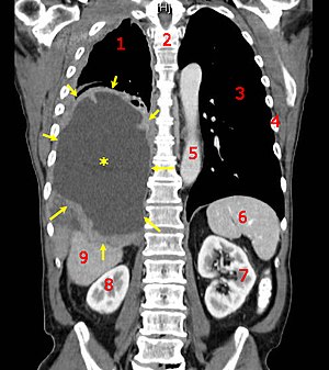 CT scan of a patient with mesothelioma,coronal section(the section follows the plane the divides the body in a front and a back half). The mesothelioma is indicated by yellow arrows, the centralpleural effusion(fluid collection) is marked with a yellow star. Red numbers: (1) right lung, (2) spine, (3) left lung, (4) ribs, (5)descendingpart of theaorta, (6)spleen, (7) leftkidney, (8) right kidney, (9)liver.>